
 Understanding points
B3.3.1 Adaptations for movement as a universal feature of living organisms (HL only)
B3.3.2 Sliding filament model of muscle contraction (HL only)
B3.3.3 Role of the protein titin and antagonistic muscles in muscle relaxation (HL only)
B3.3.4 Structure and function of motor units in skeletal muscle (HL only)
B3.3.5 Roles of skeletons as anchorage for muscles and as levers (HL only)
B3.3.6 Movement at a synovial joint (HL only)
B3.3.7 Range of motion of a joint (HL only)
B3.3.8 Internal and external intercostal muscles as an example of antagonistic muscle action to facilitate internal body movements (HL only)
B3.3.9 Reasons for locomotion (HL only)
B3.3.10 Adaptations for swimming in marine mammals (HL only) |
Muscle structure

Muscle fibre structure
Multinucleate | Muscle fibres are individual muscle cells fused together → many nuclei |
Many mitochondria | Muscle contraction requires ATP hydrolysis |
Sarcoplasmic reticulum | Specialized endoplasmic reticulum that stores Ca²⁺ ions |
Tubular myofibrils | Made up of thin actin filament and thick myosin filament |
Sarcolemma | Continuous membrane surrounding the muscle fibre that contains invaginations called T tubules |
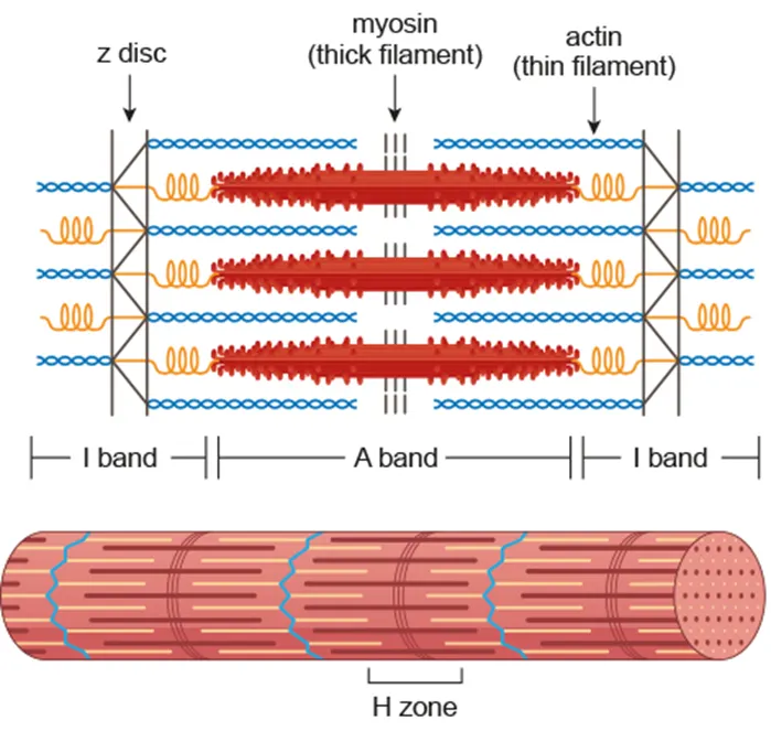
Sarcomere
Repeating contractile units composed of two myofilaments: actin and myosin
•
The thick filament (myosin) contains small protruding heads which bind to the thin filament (actin)
•
Movement of these filaments relative to one another causes lengthening /shortening of sarcomere
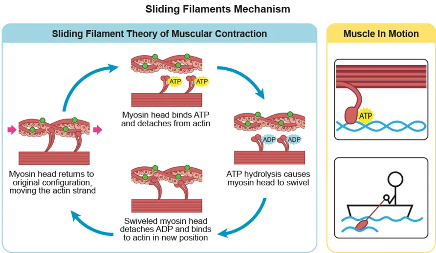
Sliding filament model of muscle contraction
Nerve impulse depolarizes sarcolemma
↓
Ca²⁺ released from sarcoplasmic reticulum
↓
Ca²⁺ bind to troponin, causing tropomyosin to expose binding sites on actin
↓
Myosin heads form cross-bridge with actin
↓
Myosin heads cause a power stroke by pulling
↓
Z-line moves closer
↓
ATP binding breaks cross-bridge = moves back to original place |
Muscle relaxation: by titin
•
Stores potential energy during muscle relaxation, releases it during contraction
•
Connects myosin filaments to the Z disc and keeps them in the center
•
Prevents overstretching of the sarcomere
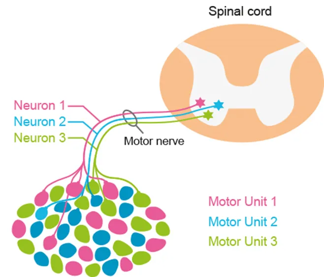
Motor units
•
Neuromuscular junction: the synapse between a motor neuron and a muscle fiber
•
Motor unit = motor neuron + all the muscle fibers it stimulates
•
Allows coordinated contraction of muscles
Intercostal muscles
•
The antagonistic action of internal and external intercostal muscles enables ventilation of the lungs
•
Internal muscles contract: ribcage moves in and down → lung volume decreases → exhalation
•
External muscles contract: ribcage moves up and out → lung volume increases → inhalation


Synovial joints
•
Capsules that surround the articulating surfaces of two bones
•
Provide structural stability by allowing certain movements
•
Composition:
1.
Joint capsule : seals joint space and provides stability by restricting the range of movement
2.
Cartilage: lines the bone surface to facilitate smoother movement, absorbs shock, distributes load
3.
Synovial fluid: provides oxygen and nutrition to the cartilage, as well as lubrication
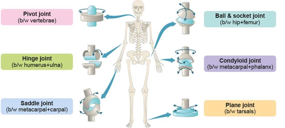
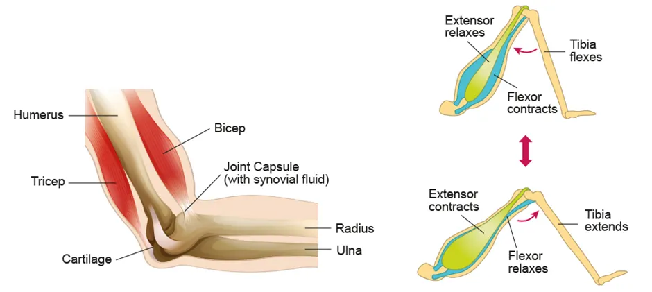
Human elbow
•
A hinge joint that is located between the humerus and radius / ulna
•
Capable of angular movement in one direction
•
Biceps : bends forearm (flexion)
•
Triceps : straightens forearm (extension)